


Reconstructive Surgery Seattle / Bellevue – Pratt Plastic Surgery
Serving Patients in Tacoma, Everett, Seattle and Bellevue
Traumatic Injury Plastic Surgery
Without a doubt, the highly skilled Seattle plastic surgeon Dr. David F. Pratt has proven himself to be a world-class cosmetic plastic surgeon. However, some of the most satisfying, memorable, and appreciated procedures he has performed have not been of the “nip and tuck” variety, but surgeries that have helped those unfortunate enough to suffer devastating traumatic injuries both during work and recreational activities. In addition to the performing incredible transformations of people’s faces and bodies through cosmetic surgery, Dr. Pratt has worked thousands of hours while covering emergency call for hospitals at which he served on the surgical staff. Often working on weekends and deep into the night, Dr. Pratt has painstakingly repaired the damage caused to faces and hands of both adults and children by falls, dog bites, and power saw injuries, auto, motorcycle, bicycle, and boat accidents.
Dr. Pratt has performed hundreds of microsurgical procedures, repairing the vital nerves, arteries, and veins of the hand and fingers of those unfortunate enough to suffer injuries while working with knives and power saws. Fingertip injuries are one of the most common injuries to occur in the hand. Dr. Pratt has utilized his skills in preserving the length and function of hundreds of fingers following traumatic fingertip injuries and amputations by performing “flap operations” in which a portion of the damaged finger or adjacent parts of the hand are utilized in “rebuilding” the traumatized finger(s).
Although Dr. Pratt did not invent these procedures, he certainly has perfected their use over the years. View actual trauma and reconstructive surgery patients who have been cared for by Dr. Pratt.
High velocity facial injuries such as those occurring as a result of falls from great heights often result in severe facial fractures, including fractures involving the eye socket.
Dr. Pratt’s specialized training in Plastic and Reconstructive Surgery has prepared him to deal with these complex injuries. In dealing on a regular basis with these types of injuries, Dr. Pratt has attained immeasurable insight and knowledge regarding the anatomy and function of the face, which has proven to be invaluable to him while performing facial cosmetic procedures.
The following cases represent just a sampling of these life changing procedures performed by Dr. Pratt over the past fourteen years.
CAUTION: Surgically Graphic Images Below
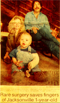
Patient #1:A two year old boy sustained a severe axe injury to his left hand resulting in complete amputation of the middle finger and severe injury to the ring finger.
The patient was brought to surgery by Drs. David F. Pratt (Resident in Plastic Surgery) and Toby Meltzer (Attending Physician) at the Oregon Health Sciences University in Portland where the amputated finger was successfully reattached and the injuries of the other finger repaired during the six hour operation.
This surgical procedure represented the first successful finger reattachment in a two year old patient ever in the state of Oregon. This patient went on to have near full use of his injured hand.
 |
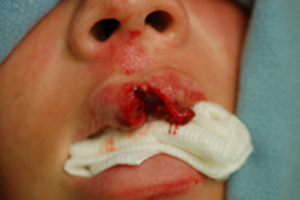
Patient #2:A sixteen year old girl fell off of her bicycle and struck her upper lip on a rock on the side of the road. She sustained a full thickness wound with loss of skin and muscle tissue and is seen here with irregular wound edges and ground in dirt. Dr. Pratt performed a single emergency reconstructive surgery on the day of the injury. The patient is seen here six weeks following her surgery.
 |
| View complete before and after images |
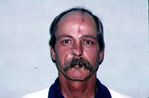
Patient #3:A forty two year old man was treated for a squamous cell cancer of the skin of his nose with removal of the tumor and the surrounding margin of skin, resulting in loss of the majority of the right side of his nose.
Dr. Pratt performed reconstruction of the defect using cartilage grafts from the ear and a “forehead flap” to provide skin coverage over the new cartilage skeleton. The skin of the forehead was elevated and the blood supply maintained via a vascular pedicle extending from the brow bone upward towards the hairline.
The “forehead flap” is rotated down to the nasal defect on this vascular leash and sutured in place. Once the new blood supply is established at the nasal recipient site two weeks later, the flap is divided and inset.
 |
| View complete before and after images |
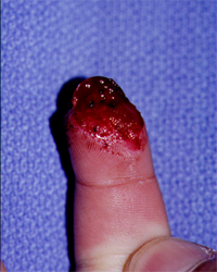
Patient #4:A sixteen year old young man sustained amputation of the tip of his dominant right index finger when it was crushed between a bicycle sprocket and chain.
The patient wished to maintain the length of the finger, thus the adjacent middle finger was attached to the index finger and skin from the middle finger was used to cover the index finger wound (called a cross finger flap) from the adjacent middle finger was used to cover the defect and a skin graft was used to cover the resultant defect of the middle finger donor site.
The fingers were surgically separated two weeks following the original flap surgery. The patient is seen here six months following his injury, fully healed with normal range of movement and excellent at the tip.
 |
| View complete before and after images |
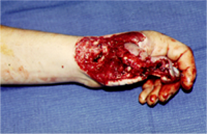
Patient #5:A twenty three year old gentleman sustained a severe power saw injury to his dominant right thumb with a Skil saw resulting in near amputation of the thumb, with actual loss of the thumb metacarpal bone, and irreparable damage to tendon, arteries and nerves.
Dr. Pratt reconstructed the patient’s thumb by taking tissue harvested from the opposite forearm including part of the radius bone, tendon, nerve, artery, and vein as well as skin and subcutaneous fat which was brought to the injured thumb and was used to reconstruct the damaged structures.
The blood supply of this so-called “free flap”, including the veins and artery connected to the blood vessels of the damaged hand. The metacarpal bone was replaced with the “free flap segment of radius bone from the opposite wrist.
After healing from this operation, the patient underwent two additional “revision surgeries” aimed at improving the alignment of the metacarpal bone and thinning out the soft tissue of the back of the thumb.
The patient underwent many months of intensive hand therapy in order to learn how to use his reconstructed hand. The patient is seen here approximately eight months following his injury. He has near normal function including excellent feeling in the skin of the thumb.
 |
| View complete before and after images |
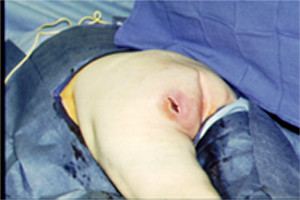
Patient #6: A sixty four year old man who suffered a stroke and was sent to a nursing home to rehabilitate developed a “pressure sore” in the buttock region. Attempts were made to heal the wound with salves and dressing changes however the wound progressed to a deep hole with bone infection at its base.
Dr. Pratt surgically removed the dying tissue and then performed a “muscle flap” procedure, using a muscle from the inner thigh to fill the wound defect and help heal this chronic pressure sore.
The patient is seen here three months following his surgery with a well healed wound.
 |
| View complete before and after images |
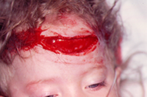
Patient #7: A three year old girl banged her forehead on a book shelf and sustained a large laceration. Dr. Pratt repaired the laceration in the Emergency Room under intravenous sedation. The patient is seen thirteen months following her injury.
 |
| View complete before and after images |
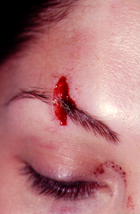
Patient #8:A twenty three year old woman struck her brow on a door and sustained a laceration through the eyebrow. Dr. Pratt repaired the laceration and the patient is seen here 1 year following laceration repair.
 |
| View complete before and after images |
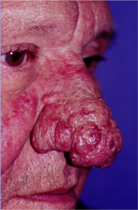
Patient #9:A fifty four year old man suffered from Rosacea for many years and eventually developed a severe rhinophyma or severe glandular enlargement of the nasal surface. Dr. Pratt performed laser excision of the excess glandular tissue and the patient is seen here six months following surgery.
 |
| View complete before and after images |
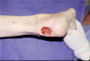
Patient #10:A sixty six year old gentleman who developed a non-healing sore on the back of his heal. Previous skin graft surgery by another physician was unsuccessful, thus Dr. Pratt performed a “neurovascular pedicle flap” which borrowed skin and subcutaneous tissue from the side of the ankle to patch the open wound on the heel. The patient is seen here eight months following surgery, completely healed and walking with a cane.
 |
| View complete before and after images |